 I’m a Nuclear Medicine Technologist, and I have been performing lung imaging here at Royal Jubilee Hospital (RJH) since 1981. Over the years, I have seen several different ways of conducting imaging that shows air supply to a patient’s lungs.
There are many reasons for doing a lung scan, but the two most common are: pulmonary embolism (this happens when a blood clot gets stuck in an artery in the lung, blocking blood flow to part of the lung), and surgical planning for lung cancer. In both cases, the patient can be short of breath, and many come to us with chronic obstructive pulmonary disease (COPD), which refers to a group of diseases that cause airflow blockage and breathing-related problems.
Past imaging technology like Xenon resulted in image quality that was hit and miss. The patient had to be able to take a good deep breath and hold it for as long as they could in order for us to image them. If they were unable to take a deep breath, the gas would not get fill their lungs enough, resulting in very poor images. The same challenge occurred if the patient could not hold their breath for more than a few seconds. This technology also only allowed technologists to capture a posterior view, which was very limiting.
Later, nebulized radioactive mist was a great improvement for imaging, but it could be just as problematic for patients. The process had patients breathing on a closed system with nose clamps on, and either a mask or a mouthpiece. This was a lengthy process, taking up to six minutes to complete, which some patients found exhausting. If the patient was short of breath, or uncomfortable with the mouthpiece or nasal clamp, we occasionally had to stop and start again. Imaging was better, but the issues with this system are many, and could obscure some details of the patient’s air supply.
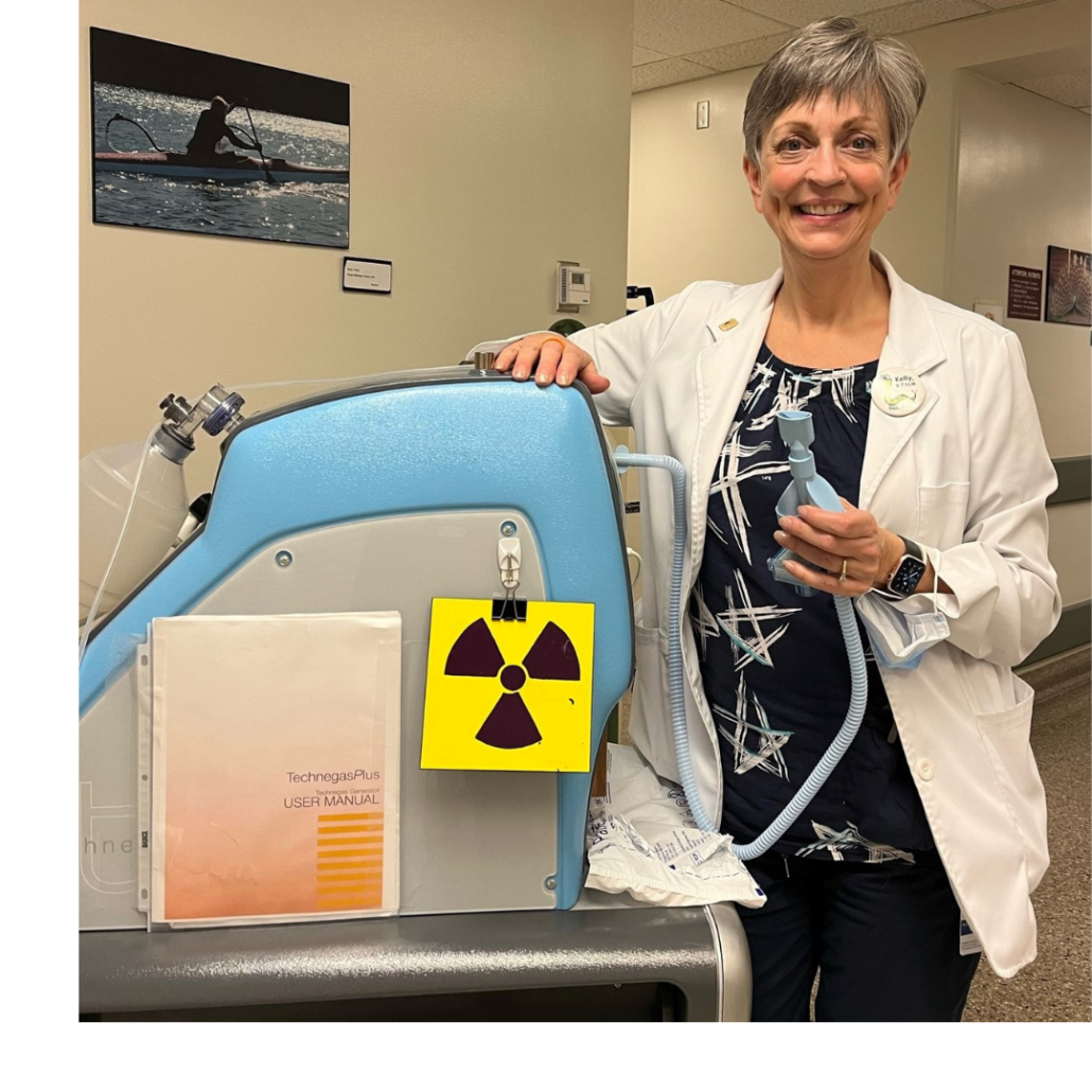
Now, I am grateful to share that, thanks to a generous $20,000 donation from the TB Vets Charitable Foundation through the Victoria Hospitals Foundation, a new Technegas Lung Imaging System arrived at RJH’s Nuclear Medicine department in 2022.
I LOVE this system. It provides beautiful images with only one breath—and occasionally two breaths—which is much easier and more comfortable for the patient. This reduces the gas delivery time from upwards of five minutes down to 20 seconds. The images only show the air supply to the lungs, and not the mouth, esophagus, or stomach as in past radioactive nebulized imaging. In short, this system provides stellar imaging with minimum stress to the patient, and in turn, is much simpler for the technologist. This system also allows us to get such clear images that we can actually get 3D imaging of the lungs, vastly improving what radiologists are able to report.
We also have a new category of lung investigation in recent years, which is called chronic thromboembolic pulmonary hypertension (CTEPH). This occurs when there is elevated blood pressure in the pulmonary arteries caused by chronic blood clots, which obstruct the free flow of blood through the lungs. These folks are often extremely short of breath, again making this new Technegas system a godsend—we can get extremely good images even if the patient isn’t able to take a deep breath.
In short, the Technegas Lung Imaging System upgrade has truly meant better service for our patients and incredibly improved imaging. Many thanks to TB Vets for helping to fund this equipment for our department.
—Kelly Norman, RTNM, Royal Jubilee Hospital
 Since 1982, TB Vets Charitable Foundation has funded an outstanding $1.5 million of urgently needed respiratory equipment at Royal Jubilee and Victoria General hospitals, helping countless patients—from newborns to seniors—receive state-of-the-art respiratory care. The Victoria Hospitals Foundation is incredibly grateful for this long-standing partnership with TB Vets.
The TB Vets Charitable Foundation supports respiratory needs throughout the province. Funds are primarily raised through the TB Vets Key Tag program, started in 1946 to support vital research, equipment, and education.


